Introduction to the Excretory System:
The human excretory system plays a vital function in preserving general health by facilitating the body’s waste disposal. This system consists of vital organs like the kidneys, bladder, and intestines, each of which makes a distinct contribution to the removal of waste. It closely cooperates with the digestive system, which breaks down food and gets waste ready to be expelled. Urine, which is produced by the kidneys filtering toxic waste products from the blood, is held in the bladder until it is released through the urethra.
The excretory system is involved removing nitrogenous wastes from body. It also regulates the amount of water and ions present in the body fluids.
Organs of Excretion in animals:
Contractile Vacuoles in Paramecium
A contractile vacuole of Paramecium is essential for osmoregulation. Excess water that enters the Paramecium is collected by the contractile vacuole, which periodically contracts to release the water from the cell. This lessens the chance that the cell will enlarge and possibly explode from the water inflow. The paramecium’s ability to maintain homeostasis and survive in its aquatic environment depends on its ability to contract and expel water.
Protonephridia in flatworms
Protonephridia composed of branched tubules that empty wastes through excretory pores on their surface. The protonephridia contain numerous flame cells with clustered, beating cilia that propel fluid into the tubules. These structures function in waste excretion and osmotic regulation.
Nephridia in earthworm
Earthworms have nephridia for excretion of nitrogenous wastes. Each nephridium consists of a tubule with ciliated opening (nephrostome) on one end and an excretory pore (nephridiopore) that opens to the outside of the body at the other end. Fluid is moved in by cilia. Some substances and water are reabsorbed in a network of capillaries that surround the tubule.
Malpighian Tubules
The excretory organs of insects are malpighian tubules. From the surrounding hemolymph (blood), they draw water and uric acid, which they then expel into the stomach. Waste products stay in the intestine while water and beneficial substances are reabsorbed.
Kidneys in Vertebrates
The kidneys of vertebrates function in the removal of nitrogenous and other wastes and in osmotic regulation of the body fluids.
Structure and Anatomy of the Kidney:
Kidneys are surrounded by adipose tissue. The cortex is where the blood is filtered. The medulla contains the collecting ducts which carry filtrate to the pelvis. Urine collects in the pelvis, a hollow cavity, and then exits into the ureter. The anatomical and functional components of the kidney are called nephrons.
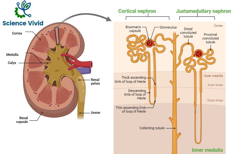
Fig: Anatomy of Kidney and Structure of Nephron
Nephron- Functional Unit of the Kidney:
Renal corpuscle
Renal corpuscle is filtering component, consisting of glomerulus and Bowmann‘s capsule. An afferent arteriole enters the glomerulus and an efferent arteriole leaves it. Bowman’s capsule is a funnel-like structure that collects filtrate from the glomerulus. The glomerulus is composed of a capillary tuft, that receives blood from an afferent arteriole. A capillary tuft that accepts blood from an afferent arteriole makes up the glomerulus. A capillary tuft called a glomerulus is where fluid exits the circulatory system. Bowmann’s capsule, which consists of an outer layer called parietal and an interior layer called visceral, envelops the tuft. Primary urine is formed in the area between two layers. The parietal outer layer is composed of simple squamous epithelium. The visceral layer is composed of podocytes. There is an ultrafitration of blood plasma. The filtration bariier is endothelium, podocytes and their basement membranes. The glomerular blood pressure provides the driving force for blood plasma to be filtered out into the space made by Bowman’s capsule. Filtration is assisted by three pressures: Glomerular capillary pressure( A positive pressure of 50-70 mm Hg exerted by blood coming from afferent artery into glomerular capillary), Colloidal osmotic pressure (A negative pressure of 30 mm Hg exerted by protein), Capsular hydrostatic pressure (A negative pressure of 15 mm Hg exerted by the filtrate already present in Bowman’s space). Net filtration pressure of 10 mm Hg. About 19% of total blood flow is Glomerular filtrate.
Renal tubules
The proximal tubule, loop of Henle, distal tubule, and collecting tubules make up the renal tubule, which is specialized for reabsorption and secretion. The proximal tubule leads from the Bowman’s capsule to the Loop of Henle. It is lined by simple cuboidal epithelium. Selective reabsorption occurs in the proximal convoluted tubule. Glucose, vitamins, important ions and most amino acids are reabsorbed from the tubule back into the capillaries near the proximal convoluted tubule. Cells of the proximal convoluted tubule have numerous microvilli and mitochondria which provide surface area and energy.
It is the most active site for reabsorption. Glucose, lactate, amino acids (almost 100%) are reabsorbed. K+(55%), Ca++, Na+ and H2O (65%) Cl–(50%), HCO3–(90%) are reabsorbed. Na+ reabsorption from lumen occur by primary active transport by basolateral Na+ K+ ATPase. Glucose, Amino acids, ions, Vitamins are reabsorbed by secondary transport mechanism. Water, urea and fat soluble substances are reabsorbed from lumen to blood by diffusion. Water is reabsorbed by paracellular route.
Loop of Henle
The loop of Henle is a long loop which extends into the medulla. Descending limb of loop of Henle is made of squamous (permeable to water). Water moves out of the descending loop as it passes through the area of high salt concentration produced by the ascending loop. The descending loop is not permeable to ions.
Ascending limb of loop of Henle is made of cuboidal (transport of ions). In the ascending loop, salt is actively pumped out. Water re-entry is not possible through this portion of the loop. As a result, a concentration gradient is produced, with the medulla (inner area) having the highest concentration.
Distal Convoluted Tubule (DCT)
The distal tubule connects the loop of Henle to the collecting duct. Simple cuboidal cells line the distal tubule. Early distal tubule is impermeable to water. Na+(5%) is reabsorbed by Na+– Cl symporter. Ca++ reabsorption (by Ca++ channel) is regulated by Parathormone hormone (PTH) and Calcitonin. In late DCT and Collecting tubule(CT), Na+ reabsorption is dependent on Aldosterone. At the same time, K+ is excreted. Water reabsorption is dependent on Antidiuretic Hormone (ADH).
Some wastes are actively secreted into the fluid in the distal convoluted tubule by a process called tubular secretion. Some of these are H+, K+, NH4+ toxic substances and foreign substances (drugs, penicillin, uric acid, creatinine). Secretion of H+ adjusts the pH of the blood.
Collecting Duct
Several renal tubules drain into a common collecting duct. The collecting ducts pass through the concentration gradient that was established by the loops of Henle. As fluid passes through the collecting ducts, much of the water moves out due to osmosis. The permeability of the collecting duct to water is regulated by hormones.