Introduction:
Microscopy is a technique of using the microscope. Microscopes are used to observe structures below the range of magnifies lens. A microscope is an optical instrument consisting of a lens or a combination of lens for obtaining magnified images. The main function of a microscope is to increase the magnification as well as to show the contrast and resolution. Magnification may be defined as the ability of lens to enlarge the image of objectives. Magnification by microscope is the product of the ocular and objective lenses. For example, if the ocular is 10X and the objective is 10X, the specimen is magnified 100 times.
The study and development of microbiology had not been possible without microscope because visual observation of microbes was made possible by the use of microscopes.
The invention of light microscope in the Sixteenth century is the single most important contribution ever made to the advancement of biology. Ancient Romans, Greeks and Indians had known lenses of glass and quartz. In the 14th century spectacles and single lenses were used as magnifiers. Campini, an Italian scientist, found that lens can be grouped to the desired curvature. Since the curvature of the lens increases its magnifying power, thereafter lens become a powerful tool in the hands of biologists. Leeuwenhoek constructed a simple, single lens microscope in 1600 AD. In 1665, Robert Hooke, an Englishman, constructed the first compound microscope, that could be truly referred to as forerunner of the modern-day microscope.
Resolving Power:
It is defined as the ability of lenses to distinguish between two smaller objects which are situated very close to each other. The resolving power of a microscope depends on two factors- the wavelength of the light used for illumination and the numerical aperture (NA) of the objectives.
Types:
Based on the principle on the magnification, modern microscopes are of two types, namely light microscope and electron. In a light microscope, magnification is obtained by a system of optical lenses using light waves, while in electron microscope the magnification is obtained by a system of magnetic field using a beam of electrons.
Light microscopes
- Bright filed microscope
- Phase contrast microscope
- Dark filed microscope
- Fluorescence microscope
- Interference microscope
Electron microscope
- Transmission electron microscope (TEM)
- Scanning electron microscope (SEM)
Light microscopes:
Bright filed (Light) Microscope
The bright filed or light microscope are the most common instruments in the microbiology laboratory. In brought filed microscope, the microscopic filed (the area observed) is brightly lightened and microorganism appear dark because they absorb some of the light. Generally, microorganism do not absorb much light, but staining them with a stain generally increases these light absorbing abilities which results in much contrast and colour differentiation.
In general, light microscopes produce a useful magnification of about 1000X to 2000X. The basic limitation of light microscope is not magnification but the resolving power. Simple, compound and or dissecting microscope are the most common bright field microscopes used in biology laboratories. Generally, compound microscopes are equipped with the three different types of objectives, each capable of different degree of magnification. These objectives are oil immersion, high dry and low power objectives. The total magnification of a microscope is determined by multiplying the magnification power of the objectives and eyepiece.
Limitation of bright field microscope: Viewing living organism is not possible without staining due to which cell will die.
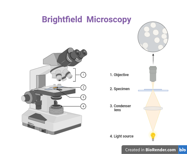
Fig: Bright filed microscope
Dark Filed Microscopy
The effect produced by the dark filed microscopic technique is bright object against a dark background. This is obtained by attaching the light microscope with a special kind of condenser-dark field condenser. In a dark filed, condenser, the center of the top lens is opaque, so that nine of the central rays of light can pass through, and the specimens on the stage is illuminated only with the peripheral oblique rays. The light rays are focused in such a way that they do not reach the specimen directly but instead the light rays objective lens. As a result, the surrounding appears dark but the specimen appears bright refractile structure.
Uses of dark field microscope: It is useful for the examination of live, unstained microorganisms or other specimens. It is also useful for the study of very sample bacteria which are generally invisible under a light microscope. Motility of microbe can be observed because microbes are in a moist, living condition.
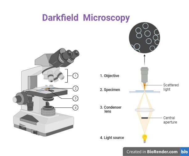
Fig: Dark field microscope
Phase contrast Microscope
The main difficulty in examining cellular organelles is that most protoplasmic materials are completely transparent.
For this reason, stained slides are usually used in bright field cytological studies. But staining of cell is resulting in cellular death, so it is natural that there some changes occur. Thus, it results in study of cellular structure which is not an original structure.
Phase contrast microscope is a special microscopic that enables the viewer to see transparent protoplasmic components without staining and thus without killing. The first phase contrast microscopic was developed in 1993 by Federick Zernike and hence it is also called Zernike microscope.
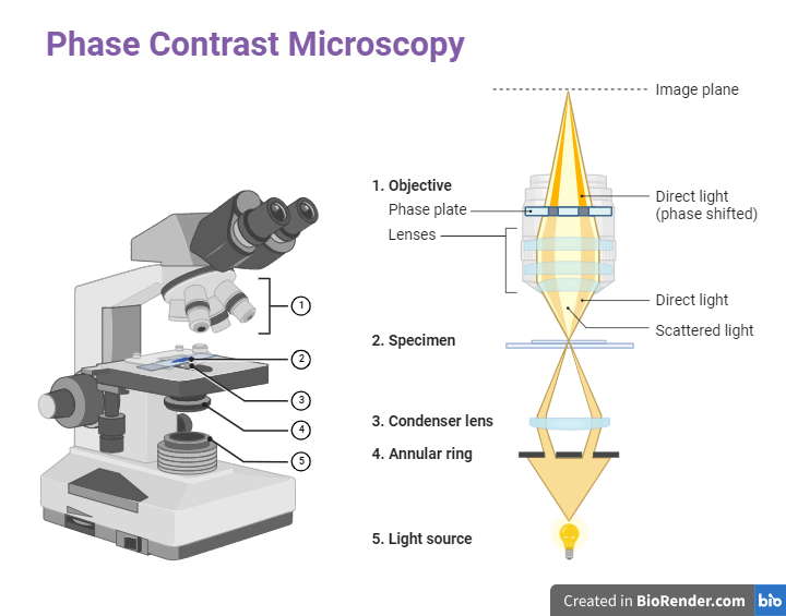
Fig: Phase contrast Microscope
Phase contrast microscope is a normal bright filed microscope, attached with a phase contrast objective and phase contrast condenser. This special attachment makes it possible to distinguish unstained structures within a cell which differ only slightly in their refractive index or thickness.
The basic idea behind this technique is that light passing through one material and into another with a slight difference in thickness will undergo a phase change. This difference in phase is translated into variation in brightness of structure and hence is detected by the viewer. As in the phase contrast microscope small phase changes results in interference of light waves resulting in high contrast images, some structures which are difficult to stain and observe under light microscope become visible.
Fluorescence Microscope
Fluoresce microscope involves staining of the specimens with fluorescent dyes. A fluoresce microscope differs from an ordinary bright field microscope in the following ways:
- It utilizes a mercury vapor arc lamp as a light source.
- Bright filed condenser is replaced by a dark filed condenser
- It employs three sets of filters to alter the light rays that pass through the instrument and reach the eye.
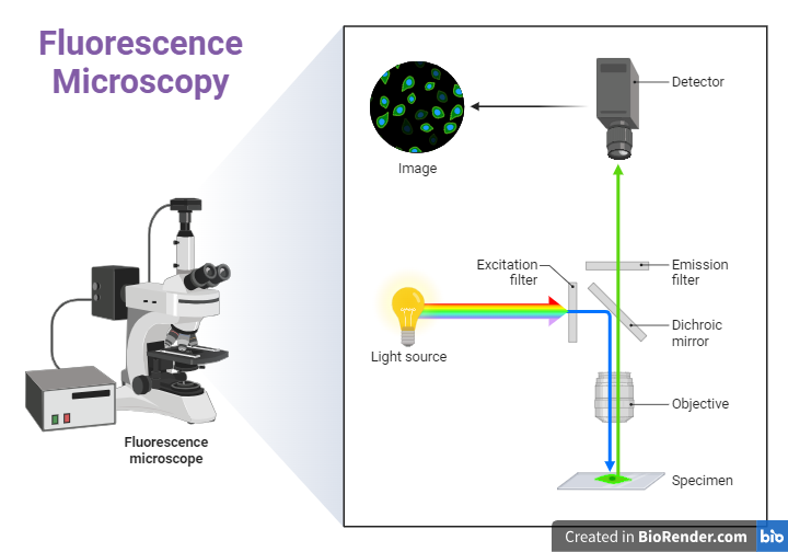
Fig: Fluorescence Microscope
In this microscope, the objects appear bright when fluorescent and the background appears light. In this microscope, UV-light is used as the exciting light and resulting fluorescent rays are always of longer wavelength than exciting rays. Microbiological materials that are studied with a fluorescence microscope must be coated with special compounds that possess this quality of fluorescence. Such compounds are called fluorochromes, for example- Auramine O, Acridine orange, Fluorescein, etc.
Interference Microscope (Stereomicroscope)
The interference microscope and phase contrast microscope operate on the concept of a change in refractive index resulting in phase contrast. In the phase contrast microscope, a single beam of light passing through the object is used, while in an interference microscope, two beams of light are used, which combine after passing through the specimen. This produces an interference pattern. Interference microscope used color to increase the contrast.
Nomarski differential interference contrast microscope is the most frequently used interference microscope. It contains polarizing filters, an interference contrast condenser and a prism analyzer plate. In the microscope, a beam of polarized light is split into two beams at right angle to each other and passed through the specimen and are combined and forced through an analyzer. The resultant interference pattern will produce a pseudo three-dimensional image, because two beams of light passing through the specimen produce a stereoscopic effect. Images seen through this microscopic are normally brilliantly colored (non-stained) because of the phase changes of light waves that pass through the various components of an object.
Electron microscope:
This microscope differs in may aspects from light microscope. The electron microscope provides tremendous magnification because of the much higher resolution obtained with the extremely short wavelength of electron beam used to magnify the specimen. Electron microscope used electron beam used to magnify the specimen. Electron microscope used electron beam and magnetic field to produce the image, while light microscope uses light waves and glass lenses to produce image.
Electron microscope employs 60 to 80 Kv electrons having wavelength of only 0.05 A0.
By unsung electron microscope it is possible to resolve objects as small as 10 A0 and it magnifies the image unto 400000 X to 1000000X.
The basic components of electron microscope are electron generator (gun), 3 sets of electromagnetic coils, screen for viewing and vacuum pump. Th electron generator is a tungsten filament, which when heated by electric current emits a stream of electrons. From there, this electron beam passes through the condenser. Condenser is magnetic coil which corrects the bending in the beam and directs it towards the object.
For electron microscope, the specimens, the specimen to be examined is prepared as an extreme thin, dry, film on small screen and introduced into the instrument at a point between the magnetic condenser and magnetic objective. This point is comparable to the stages of the light microscope. Th electron beam passes through it and is scatted depending on the varying refractive index of the specimen. From there, the electron beam passes through the sound set of magnetic coils which focus the electrons and form an intermediate image. The third set of magnetic lenses, the projection lenses, produce the final image. However, the final image cannot be directly viewed as the electron rays are harmful to the eyes. They are either projected on the fluorescent screen through the airtight window on recorded on a photographic plate by a camera built into the instrument.
Applications
- Viewing of ultra-structure of cell organelles is possible.
- Structural details of large biomolecules such as nucleic acid, protein, etc, can be studied.
- Minute details of the topographic architecture cab be studied.
Limitations
- Despite the great advantage of better resolution and higher magnification, there are several limitations to electron microscopic studies. These limitations are
- The specimens being examined in a chamber that is under a very high vacuum, the cells cannot be examined in a living state.
- The drying process may alter some morphological characteristics.
- As electrons have a low penetration power, it is necessary to use thin sections of specimens’ cells to reveal the internal structure of the cell.
- The entire electron microscope must be in a vacuum or otherwise the electron get scatted due to collisions with air molecules and thus fail to get focused. There are two main types of electron microscopes.
Transmission Electron Microscope (TEM)
Studying the structural details of the various components present in a cell can be achieved using TEM. The specimens form a contrasting image with all the structural details. Numerous techniques are available for use with TEM, which extends its usefulness in charactering cellular structure. Some of these are-shadow casting, Negative staining, Ultrathin Sectioning, Freeze etching, Localization of cell constituents, Localization of enzymes in thin sections, Autoradiography etc.
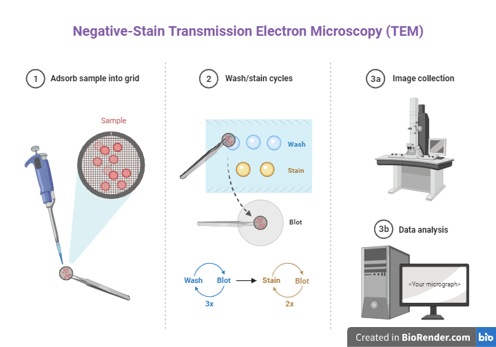
Fig: Negative-Stain Transmission Electron Microscopy (TEM)
Scanning Electrons Microscope (SEM)
SEM is primarily used for visualizing the surfaces architecture of the specimen instead if internal details. The general construction and working principle of a SEM is different from that of TEM. In SEM, that specimen is subjected to a narrow beam of electrons which rapidly move over (scan) the surfaces of that specimen. This causes the release of a shower of secondary electrons and other types of radiation from the surfaces of the specimen. The intensity of these secondary electrons depends on the shape and the chemical composition of the specimen These secondary electrons are captured by a detector, and they generate an electrical signal. These signals are then scanned in the manner of a television system to produce an image in a cathode ray tube.
The SEM lacks the resolving power obtainable with TEM, but has the advantages of revealing a striking three-dimensional picture. The surface topography of as specimen can be revealed with a clarity and a depth of filed not possible by any other method.
