Introduction to the Spinal Cord:
The spinal cord is the elongated caudal part of the CNS. It starts as the inferior continuation of the medulla oblongata at the level of foramen magnum, & ends as an inverted cone (the conus medullaris) at the lower end of L1 vertebra.
The spinal cord conveys motor signals from the brain to the spinal nerves, & sensory information from the spinal nerves to the brain. Furthermore, nerve reflexes are mediated by the spinal cord alone, independent of the brain.
Morphologically, the spinal cord resembles a long cylinder with 2 swellings in the cervical & lumbosacral regions (for the innervation of the upper & lower limbs respectively). Along its length, the spinal cord gives rise to the ventral spinal nerve rootlets antero-laterally, & dorsal rootlets postero-laterally.
External Features of the Spinal Cord:
A deep median fissure running along the cord’s anterior aspect is known as an anterior median fissure. It extends into the anterior white commissure, & is covered by the anterior spinal artery and vein, containing its sulcal (central) branches, & is filled with CSF.
A shallow median depression that runs at the back of the cord is called the posterior median sulcus. It is covered by the posterior spinal vein.
The anterior rootlets are linked to the anterolateral sulcus, a small depression that runs along the cord’s anterolateral side.
The posterior rootlets are linked to the posterolateral sulcus, a shallow indentation along the cord’s posterolateral side.
Posterior intermediate sulcus: a shallow paramedian longitudinal depression between the posterior median sulcus & the posterolateral sulcus. It separates gracile fascicle from cuneate fascicle.
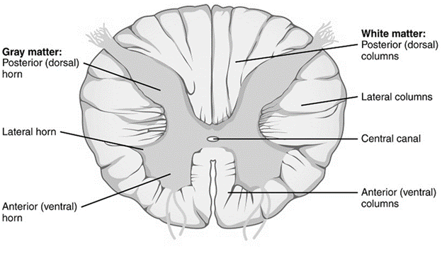
Fig: Cross-section of spinal cord
Internal Structure of the Spinal Cord:
The spinal cord is composed of an inner mass of nerve cell bodies (gray matter) encircled by a vast number of myelinated nerve fibers (white matter).
Gray Matter of the Spinal Cord
Grey matter extends along the spinal cord, & has “H” or butterfly shape in horizontal sections. Spinal cord gray matter has the following parts:
The anterior (ventral) horn
This is a gray matter column that runs the length of the spinal cord. It is short, broad, and does not extend to the spinal cord’s surface. Groups of massive multipolar neurons (also known as anterior horn cells) with a solely motor function make up the anterior horn. These neurons receive impulses from the motor cortex of the brain & send their axons through the anterior rootlets of the spinal nerves, to supply the skeletal muscles of the whole body (except the muscles supplied by the cranial nerves).
Posterior (dorsal) horn
This column of gray matter extends throughout the whole spinal cord. It is narrow, long, & almost reaches to the surface of the spinal cord. The posterior horn is made of groups of intermediate & small multipolar neurons which are purely sensory in function. These neurons receive sensory impulses from the body (through the central processes of pseudounipolar neurons in the dorsal root ganglia) & send their axons contralaterally to the higher sensory areas of the brain.
Intermediate gray zone: this is the area of gray matter existing between the anterior horn, posterior horn, & the gray commissure (the horizontal bar of gray matter). It contains neurons of variable sizes that are distributed homogenously.
Gray commissure
It is the thin transverse strip of gray matter extending from right to left, & contains the central canal of the spinal cord. The CSF-filled central canal is continuous superiorly with the 4th ventricle of the brainstem, & closes inferiorly with the end of the spinal cord.
The shape of the spinal cord grey mater differs according to each spinal cord region. Also, the proportion of the grey mater relative to the white mater increases as it descends towards the spinal cord end.
White Matter of the Spinal Cord
The white matter is located externally, & is made of bundles of myelinated nerve fibers running up & down in the cord. Grossly, the white matter is divided by the gray matter into 3 this columns (funiculi):
Anterior column (funiculus)
This is the white mater part existing between the anterior median fissure, the anterior gray horn, & the origin of the anterior rootlets. Just anterior to the gray commissure, the 2 anterior columns communicate with each other via the anterior white commissure.
Lateral column (funiculus)
This is the white mater part existing between the anterior & posterior gray horns (between the origins of the anterior & posterior rootlets).
Posterior column (funiculus)
This is the white mater part existing between the midline & the posterior gray horn. The right & left posterior columns are separated by a midline sulcus & septum (the posterior median sulcus & septum). Each posterior column is subdivided into a medial fascicle (fasciculus gracilis) & a lateral fascicle (fasciculus cuneatus), separated by the posterior intermediate sulcus & septum.
Spinal Cord Reflexes:
A reflex is an involuntary motor response to a sudden sensory stimulus. Its occurrence depends on the integrity of the reflex arc (pathway). Simply, a reflex arc consists of the following anatomical structures: (1) a receptor organ, (2) an afferent neuron, (3) an efferent neuron, and (4) an effector organ. A reflex arc involving only one synapse is a monosynaptic reflex arc.
Interruption of the reflex arc at any point along its course would abolish the response. The neurons in the reflex arc receive descending fibers from higher CNS centers (namely: the brainstem & cerebral cortex) that can modulate the reflex activity. Thus, a lesion involving those higher centers may result in abnormal reflex activity.
In the spinal cord, reflex arcs play an important role in maintaining muscle tone, which is the basis for body posture. The receptor organ is situated in the skin, muscle, or tendon (a sensory receptor).