Background:
Gram staining is a key technique in microbiology that allows bacteria to be classified into two groups: Gram-positive and Gram-negative. This staining method, developed by Hans Christian Gram in the late 1800s, gives vital insights into bacterial cell wall composition and has become a cornerstone of bacterial identification and classification. The differential staining is based on Gram-positive bacteria’s ability to hold the crystal violet dye due to their thick peptidoglycan coating, which gives the purple colour. Gram-negative bacteria, on the other hand, have a weaker peptidoglycan layer and an outer membrane that prevents crystal violet retention, resulting in a pink or red colour when counterstained with safranin.
Purpose:
Gram staining is used quickly and effectively accurately separate bacteria into Gram-positive and Gram-negative groups based on the composition of their cell walls. This understanding is useful for the initial identification of bacteria, for determining antibiotic susceptibility, and for identifying bacterial pathophysiology.
Materials:
- Bacterial culture
- Glass slides
- Crystal violet stain
- Iodine solution (Gram’s iodine)
- Ethanol or acetone (decolorizer)
- Safranin counterstain
- Water (for rinsing)
- Blotting paper or paper towels
- Immersion oil (for oil immersion microscopy)
- Microscope slide coverslips
Equipment:
- Bunsen burner or heat source
- Inoculating loop or sterile swab
- Microscope
- Slide rack or staining tray
- Pipettes or droppers for dispensing reagents
- Microscope slide holder or stage clips
- Safety goggles
- Gloves
- Lab coat or protective clothing
- Timer or stopwatch
Procedure:
- Prepare a clean glass slide by labelling it with the appropriate sample identification.
- Using a sterilized inoculating loop, transfer a small amount of the bacterial culture onto the centre of the slide.
- Use the loop to spread the bacterial culture into a thin, even film on the slide.
- Allow the slide to air dry completely.
- Pass the slide through the flame of a Bunsen burner or heat source 2-3 times to fix the bacteria to the slide.
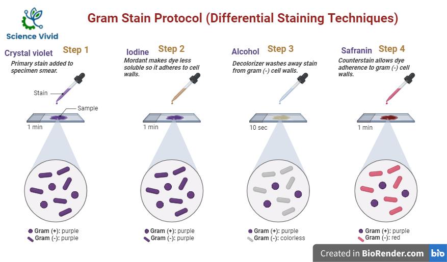
- Once the slide is cool, cover the bacterial smear with crystal violet stain and let it sit for 1 minute.

Fig: Protocol for gram staining
- Rinse the slide gently with water to remove the excess crystal violet.
- Cover the slide with iodine solution and let it sit for 1 minute. Iodine acts as a mordant to enhance the staining.
- Rinse the slide gently with water to remove the excess iodine.
- Holding the slide at a slight angle, gently pour ethanol or acetone over the slide to decolorize it. Allow the decolourizer to flow until no more colour is being washed off (usually 10-30 seconds).
- Immediately rinse the slide with water to stop the decolorization process.
- Cover the slide with safranin stain and let it sit for 1 minute. Safranin acts as a counterstain.
- Rinse the slide gently with water to remove the excess safranin.
- Blot the slide gently with blotting paper or a paper towel to remove excess water.
- Allow the slide to air dry completely.
- Once the slide is dry, it is ready for microscopic examination.
- Place the slide on the microscope stage and observe under oil immersion objective.
- Gram-positive bacteria will appear purple, while Gram-negative bacteria will appear pink or red.
Results:
- Gram staining provides visual results that help distinguish between Gram-positive and Gram-negative bacteria.
- After Gram staining, Gram-positive bacteria appear purple or blue under the microscope.
- Gram-negative bacteria appear pink or red under the microscope after Gram staining.
Interpretation:
The analysis of Gram staining results provides essential information into the cell wall composition and classification of bacteria. The interpretation of Gram staining is crucial in bacterial identification, providing preliminary categorization information and directing clinical diagnosis and treatment options. However, it is crucial to highlight that additional confirmatory test are frequently necessary for correct identification and characterization of bacteria.
Troubleshooting
When troubleshooting Gram staining, it is critical to address difficulties such as insufficient staining, overstaining, improper decolorization, poor quality of staining reagents, bacterial culture problems, slide preparation, and microscope-related concerns.
References:
- Tripathi, N. and Sapra, A., 2020. Gram staining.
- Moyes, R.B., Reynolds, J. and Breakwell, D.P., 2009. Differential staining of bacteria: gram stain. Current Protocols in Microbiology, 15(1), pp.A-3C.
- Thairu, Y., Nasir, I.A. and Usman, Y., 2014. Laboratory perspective of gram staining and its significance in investigations of infectious diseases. Sub-Saharan African Journal of Medicine, 1(4), p.168.
