Introduction:
Chromosomes are thread-like structures that are found in the nucleus of both animal and plant cells. Each cell’s nucleus contains chromosomes that bundle the DNA molecule. DNA, a type of nucleic acid, is tightly wrapped around proteins called histones that stabilize the organization of each chromosome.
History:
In the late 1800s, scientists using a microscope to examine cells discovered chromosomes for the first time. The nature and function of these cell structures, however, were unknown at the time.
Strasburger (1815) was the first to define chromosomes, while Waldeyer (1888) was the first to adopt the name “chromosome.” The Greek words “chroma” and “body” are combined to get the word “chromosome” (soma). Because chromosomes are cell structures, or bodies, that are intensely stained by several bright dyes employed in study, scientists gave them this name.
When cells are stained with an appropriate basic dye and observed under a light microscope during the metaphase phase of mitosis, they appear as rod-shaped dark stained bodies.
Thomas Hunt Morgan’s pioneering studies in the early 1900s gave researchers a far better grasp of chromosomes. Morgan established the link between chromosomes and hereditary features in fruit flies by proving that the X chromosome is linked to gender and eye colour.
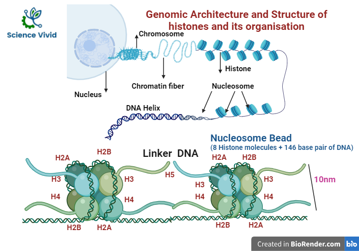
Fig: Genomic Architecture and structure of histones and its organisation
Do all living things share the same chromosomal types?
- Among the major differences between viruses, prokaryotes, and eukaryotes is the shape and organization of chromosomes. The chromosomes of non-living viruses are made up of either DNA (deoxyribonucleic acid) or RNA (ribonucleic acid), which is packed very densely inside the viral head. Chromosomes are totally made up of DNA in prokaryotic species (such as bacteria and blue-green algae). A prokaryotic cell’s single chromosome isn’t protected by a nuclear membrane. The chromosomes are placed in the nucleus of eukaryotes, which is a membrane-bound cell. DNA is coupled to a protein core in the chromosomes of eukaryotic cells. RNA is present as well.
- The number and form of chromosomes differs from one live organism to the next. One or two circular chromosomes can be found in most bacteria. Humans, like other animals and plants, have linear chromosomes organized in pairs within the cell nucleus.
- The only human cells that don’t have two copies of each chromosome are reproductive cells, or gametes. When two reproductive cells combine, a single cell with two copies of each chromosome is formed. This cell divides, and its descendants divide, generating a mature individual with a complete set of paired chromosomes in nearly all of its cells.
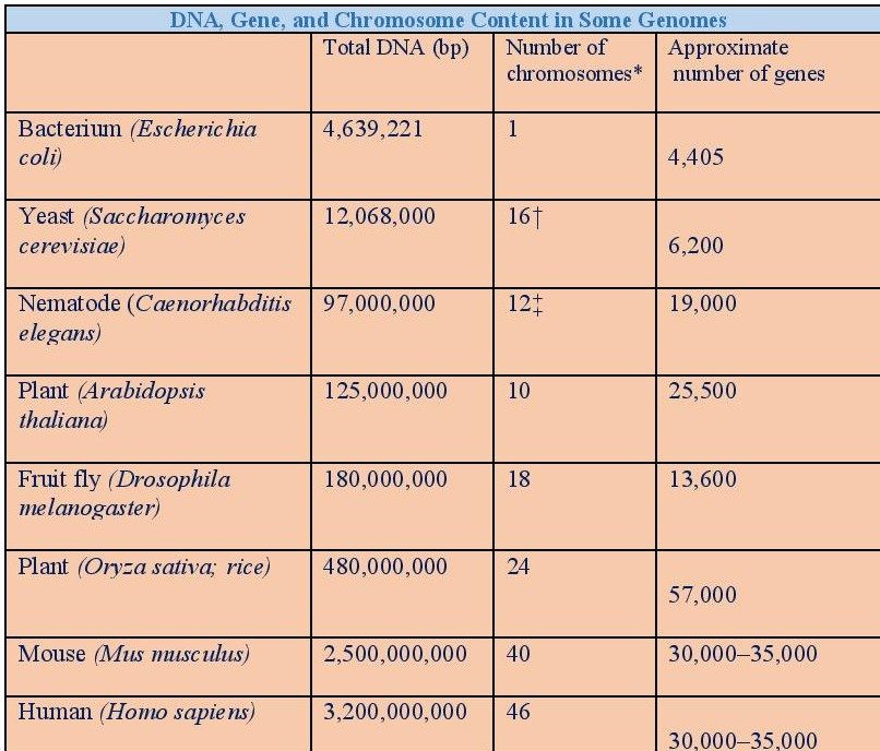
- Hundreds or thousands of genes are found on each chromosome. Within the cell, chromosomes are found in pairs. Each cell has two copies of each gene (alleles). Humans have 46 chromosomes (23 pairs), cats have 38 chromosomes (19 pairs), while dogs have 78 (39 pairs).
Note: This information is constantly being refined.
* Except for yeast, all eukaryotes have a diploid chromosomal number.
†Haploid chromosome number. Eight (octoploid) or more sets of these chromosomes are found in most wild yeast strains.
‡Number for females, with two X chromosomes. Males have an X but no Y, making a total of 11 chromosomes.
- In addition, cells can have multiple types of chromosomes; for example, mitochondria in most eukaryotes and chloroplasts in plants each have their own small chromosomes. Viral chromosomes, prokaryotic chromosomes, and eukaryotic chromosomes are the example of various types of chromosomes.
- Plants, yeast, and mammals are eukaryotes (cells with nuclei), and their nuclei contain numerous large linear chromosomes. Each chromosome contains one centromere and one or two arms protruding from it, though these arms are rarely visible. Furthermore, most eukaryotes contain a small circular mitochondrial genome, and some may have extra small circular or linear cytoplasmic chromosomes.
Organisation in the Nucleus:
The nucleus is more than just a container for chromatin, RNAs, and nuclear proteins to freely move around in aqueous solution. Chromatin is a DNA-protein complex in which the mass of the protein and the nucleic acid are approximately identical. Within cells, chromatin folds into chromosomes. A single double-stranded segment of DNA and the associated packaging proteins are found on each chromosome.
The nucleosome is the smallest structural component of chromatin, and it is formed by connecting DNA and histone proteins.
Each nucleosome is made up of an octamer core made up of histones H2A, H2B, H3, and H4 (or other histone variations in rare situations) and a DNA segment that wraps around it. “Linker DNA” connects adjacent nucleosomes.
Structure:
When the chromatin fibre is condensed and coiled into a distinctive form during mitosis, the chromosome structure is most easily identified.
Only during mitosis, when a blood cell culture is stimulated with a mitogen for three days, is the chromosome structure observable. Multiple massive, linear chromosomes are found in the nucleus of eukaryotes.Each chromosome typically has one centromere and one or two arms that project from the centromere. Eukaryotes have numerous big, linear chromosomes that are located in the nucleus of the cell.
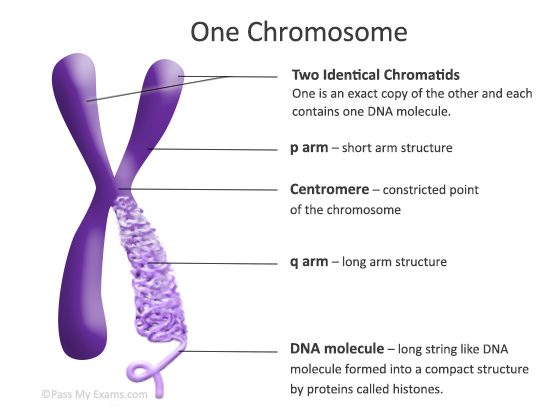
Fig: Structure of chromosome
The centromere, found on each chromosome, is a constriction point that separates the chromosome into two portions, or “arms.” The “p arm” refers to the chromosome’s short arm. The “q arm” refers to the chromosome’s long arm. The centromere’s position on each chromosome determines the chromosome’s structure and can be used to explain the location of individual genes.
Each chromosome is divided into three segments based on its structure:
- Pellicle:
It is the outer envelope around the chromosome.
- Matrix:
It is the ground substance of chromosome which also includes non-genetic materials that contain the chromonemata.
- Chromonemata:
The chromonemata is a spirally coiled threads that is embedded in the matrix of chromosome.
The following structural characteristic (apart from the chromomere) can be seen under a light microscope in mitotic metaphase chromosomes:
(1) Chromatid,
(2) Chromonema,
(3) Chromomeres,
(4) Centromere,
(5) Secondary constriction or Nucleolar organizer,
(6) Telomere and
(7) Satellite
Types:
Autosomes and Sex Chromosomes
Human chromosomes are of two types- autosomes and sex chromosomes. Sex chromosomes pass on genetic features associated to a person’s sex. The rest of the genetic information is present in the autosomes. Humans contain 46 chromosomes in their cells, with 22 pairs of autosomes and one pair of sex chromosomes.
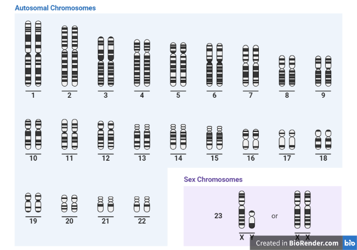
Fig: Human Karyotype
On the Basis of Number of Centromeres
- Monocentric with one centromere.
- Dicentric with two centromeres.
- Polycentric with more than two centromeres
- Acentric without centromere. These chromosomes are freshly fragmented chromosomal segments that do not survive for long.
- Diffused or non-located with indistinct centromere diffused throughout the length of chromosome.
On the Basis of Location of Centromere
- Telocentric are rod-shaped chromosomes with centromere occupying the terminal position, so that the chromosome has just one arm.
- Acrocentric are also rod-shaped chromosomes with centromere occupying a sub-terminal position. One arm is very long and the other is very short.
- Sub-metacentric chromosomes are with centromere slightly away from the mid-point so that the two arms are unequal.
- Metacentric are V-shaped chromosomes in which centromere lies in the middle of chromosome so that the two arms are almost equal.
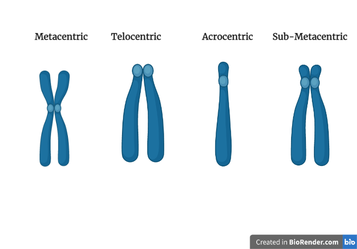
Fig: Different types of chromosomes on the basis of centromere
The Human Chromosomes:
Except for sperm and egg cells, a normal human cell has 23 pairs of chromosomes for a total of 46 chromosomes. Only one of each pair of chromosomes is present in sperm and egg cells, for a total of 23. There are hundreds to thousands of genes on each chromosome. One of the 23 chromosomal pairs are the sex chromosomes. Normal persons have two sex chromosomes, one of which is an X and the other a Y. Normal females have two X chromosomes (XX), while males have one X and one Y(XY).
Chromosome Abnormalities/Aberrations:
Cytogenetics is the study of chromosome structure and properties, including chromosomal behaviour during somatic cell division in growth and development, mitosis, germ cell division in reproduction, meiosis, chromosomal influence on phenotypes, and factors that cause chromosomal changes. Because normal chromosomes have a consistent appearance and size, chromosomal analysis provides useful information about a person’s genetic makeup. Furthermore, chromosomal abnormalities cause a variety of genetic illnesses, as well as miscarriages, implantation failures, and congenital deformities.
Chromosomal abnormalities can fall under two categories:
Structural abnormalities
When there is a change in the structure or portions of a chromosome, structural chromosomal abnormalities arise. There are 46 chromosomes in a single cell. When a component of a chromosome is missing, an additional part is present, or a part has switched positions with another section, structural chromosomal abnormalities develop. In the end, this results in an excess of or a deficiency of genetic material. Some of the birth defects are results of these abnormalities.
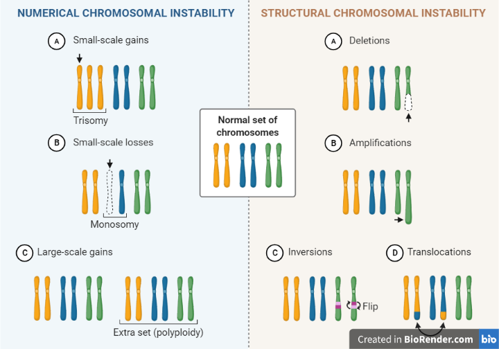
Fig: Numerical and structural abnormalities of chromosome
Numerical Abnormalities:
Numerical aberrations are chromosome defects in which the body’s cells have a different number of chromosomes than normal. As a result, each cell of the body may have 45 or 47 chromosomes instead of the usual 46.
Chromosome number abnormalities are most commonly caused by meiotic non-disjunction (the failure of chromosome pairs to split during cell division) or anaphase lag (loss of chromosome during cell division).
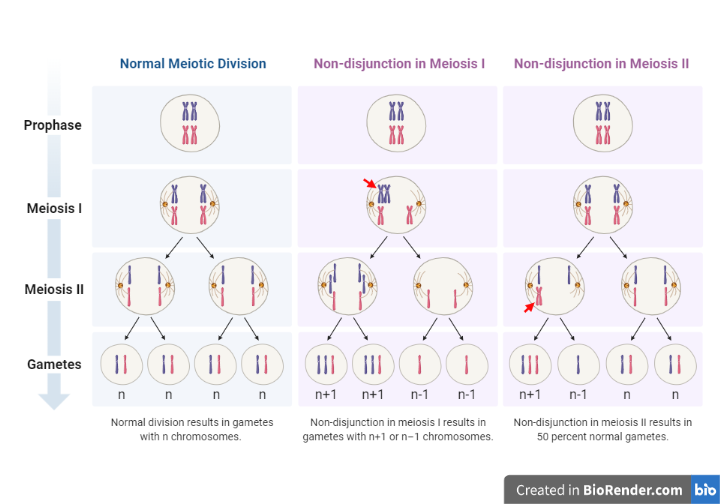
Fig: Non-disjunction in Meiosis
Risk factors for chromosomal abnormalities:
Chromosome abnormalities occur when a cell divides abnormally. In each step, the appropriate number of chromosomes should be found in the resulting cells (mitosis and meiosis). Cell division abnormalities, on the other hand, may result in cells with too few or too many copies of a chromosome.
Other factors that can raise the probability of chromosome abnormalities encompass:
Environment
Although there is no conclusive evidence that certain environmental variables induce chromosome abnormalities, it is still possible that the environment has a role in the incidence of genetic errors.
Maternal Age
Women are born with all of their eggs. Some scientists agree that as the eggs age, defects in their genetic material may occur. Older women are more likely than younger women to have children with chromosomal abnormalities. Because men produce new sperm throughout their lives, male fertility does not raise the chance of chromosomal abnormalities.
Chromosomal Disorders:
When a mutation (an undesirable change to a gene, also known as a pathogenic variation) affects the genes or have an insufficient amount of genetic material, it may result in genetic disorder. Some of the examples are:
Monogenic disorders
- Sickle cell disease
- Tay-Sachs disease
- Cystic fibrosis
- Duchenne muscular dystrophy (DMD)
Chromosomal disorders
- Turner syndrome
- Trisomy 18
- Trisomy 13
- Down syndrome (Trisomy 21)
- FragileX syndrome
- Klinefelter syndrome
- Triple-X syndrome
Multifactorial disorders
- Diabetes
- Spina bifida
- Congenital heart defects.
- Arthritis
- Autism spectrum disorder
- Coronary artery disease
Prenatal Screening for Chromosomal Abnormalities:
Prenatal genetic testing provides the information about whether the foetus has certain genetic disorders. Basically, Prenatal diagnosis of chromosomal abnormalities is currently accomplished by using blood or body fluids through invasive techniques such as amniocentesis and chorionic villus sampling to analyses the if there is any error in the chromosomes.
CVS is undertaken in the first trimester, between 10 and 13 weeks of pregnancy, whereas amniocentesis is performed at 15 weeks of pregnancy.
Likewise, the non-invasive prenatal test, or NIPT, is a new, extremely sensitive test which is used in the first trimester of pregnancy to screen for Down syndrome and certain other abnormalities in a new-born.
References:
- A.R. Cutter, J.J. Hayes / FEBS Letters 589 (2015) 2914–2922
- Ris, H. and Kubai, D.F., 1970. Chromosome structure. Annual review of genetics, 4(1), pp.263-294.
- White, P.S., 1973. Chromosome 1. eLS.
- Rizos D. (2018). Prenatal Screening for Chromosomal Abnormalities: Where do We Stand Today in Mediterranean Countries?. EJIFCC, 29(4), 274–279.
- Lehninger, A.L., Nelson, D.L., Cox, M.M. and Cox, M.M., 2005. Lehninger principles of biochemistry. Macmillan.
- Alberts, B., Bray, D., Hopkin, K., Johnson, A.D., Lewis, J., Raff, M., Roberts, K. and Walter, P., 2015. Essential cell biology. Garland Science.
- Lodish, H., Berk, A., Kaiser, C.A., Krieger, M., Scott, M.P., Bretscher, A., Ploegh, H. and Matsudaira, P., 2006. Molecular cell biology.
- Prenatal Testing for Chromosomal Abnormalities and Neural Tube Defects. ARUP Consult®. Retrieved May 31, 2022, https://arupconsult.com/content/prenatal-screening-and-diagnosis. Filoche S, Lawton B, Beard A, Dowell A, Stone P. New screen on the block: non-invasive prenatal testing for fetal chromosomal abnormalities. J Prim Health Care. 2017 Dec;9(4):248-253. doi: 10.1071/HC16055. PMID: 29530134