Introduction to the Brain:
The brain is an intricate organ that governs all bodily functions, including thought, memory, emotion, touch, motor skills, vision, respiration, temperature, hunger, and every bodily function. The central nervous system, or CNS, is made up of the brain and the spinal cord that branches off of it. The average adult’s brain weighs around 3 pounds and is composed of 60% fat. Water, protein, carbs, and salts make up the remaining 40%. The brain is not a muscle in and of itself. It has nerves, including neurons and glial cells, as well as blood vessels.
Cerebrum:
The main part of the brain, which is separated into the left and right hemispheres, is the cerebrum. The cerebrum regulates the body’s voluntary motions, such as sitting, jogging, and walking. Cerebrum is the largest part of the brain, the cerebrum, makes up roughly two-thirds of the total brain mass. It is made up of two hemispheres separated by the corpus callosum, a fissure. It consists of the basal ganglia, medullary body, and cerebral cortex. Cerebrum receives sensory impulses from the sense organs. It contains the centers of thought and memory.
Because it lacks a myelin sheath yet has cell bodies and synapses, the cerebral cortex is the part of the brain that is frequently referred to as gray matter. The grey matter, or cortex, is primarily made up of neurons and astrocytes. Because the nerves in the cortex (thin layer of tissue) do not have the white fatty myelin sheath or insulation that gives the majority of other areas of the brain its white appearance, the cortex appears gray. The outer 1.5–5 mm of the cerebrum and cerebellum are covered by the cortex.
Deep grooves or fissures known as sulci are formed by the folding bulges called gyri that make up the cortex. The brain’s surface area is increased by the folds, increasing the amount of gray matter and processing power of the brain. White matter that consists mostly of axons and oligodendrocytes. Both layers include glial cells. The white matter of the cerebrum, or medullary body, is made up of myelinated axons.
The corpus callosum is formed by commissural fibers, which carry impulses between the hemispheres. Impulses enter and exit the cerebral hemispheres through projection fibers. Association fibers conduct impulses within the hemispheres. The mass of gray matter in each hemisphere that regulates voluntary muscle movements is known as the basal ganglia.
Four Lobes of the Cerebrum
- Frontal lobe is the motor area involved in movement and in planning & coordinating behavior, memory, judgment, inhibitions, personality
- Parietal lobe is associated with sensory integration, attention, and language
- Temporal is involved in auditory perception, speech, and complex visual perceptions, long term memory
- The visual center responsible for processing visual information is the occipital lobe.
Particular areas
The frontal lobe’s Broca’s region plays a crucial role in speech production.
Wernicke’s area is important in language understanding and meaningful speech output.
The Limbic System is a collection of brain regions that help control emotional expression and emotional memory, including the aamygdala, hippocampus, septum, and basal ganglia.
Cerebellum:
Each of the hemispheres that make up the cerebellum has three lobes. The section between hemispheres is called the vermis. Cerebellum coordinates somatic motor function. Cerebellum controls fine muscle control (adjusts output of somatic motor centers resulting in smooth operation). Cerebellum maintains body balance during movement and controls aspects of motor learning.
The cerebellum consists of white matter, containing pairs of relay neurons that connect the cerebellum to other parts of the nervous system. The white matter is surrounded by a thin layer or mantle of gray matter. Within the gray matter are three layers: an outer molecular layer, an inner granular layer, and a thin layer of large Purkinje cells between the two.
There are five types of neurons in the cerebellum:
- Purkinje cells
- Stellate cells
- Basket cells
- Golgi cells
- Granule cells
Brainstem:
Brainstem connects the spinal cord to the brain. It transmits information from the spine to the upper brain and is made up of the medulla, pons, and midbrain (involuntary responses). The brainstem connects the cerebral hemispheres to the spinal cord and cerebellum. There are four separate parts of the brain stem: the diencephalon, midbrain, pons, and medulla oblongata.
Midbrain
Midbrain processes visual and auditory data. Midbrain is involved in the control of consciousness and awareness
Pons
Pons relays information to the thalamus and cerebellum. Pons regulates subconscious somatic and visceral motor centers
Medulla oblongata
Medulla relays information to the thalamus and brain stem. Medulla regulates visceral function
Medulla houses the reflex centers e.g., those associated with respiratory (breathing) and cardiovascular system (blood pressure, heartbeat), swallowing, digestion, body temperature, sleep
The ascending and descending routes between the brain and spinal cord are channeled by the medulla. The ascending and descending routes between the brain and spinal cord are channeled by the medulla.
Diencephalon
Internal organs (viscera) are controlled by the diencephalon. Diencephalon directs sense impulses throughout the body. Diencephalon control autonomic function control
Diencephalon is involved in endocrine function control, motor function control, homeostasis, hearing, vision, smell, taste and touch perception
Diencephalon has 4 divisions
- Thalamus, which is a repeating station, is in responsible of information traffic.
- Subthalamus responsible for check of muscular responses
- Hypothalamus responsible for hormone regulation, “flight and fight”, feeding and sex, and body function in general. Hypothalamus is involved in regulating activities internal organs, monitoring information from the autonomic nervous system, controlling the pituitary gland and its hormones, and regulating sleep and appetite
- Epithalamus (including the pineal gland), responsible for melatonin and the circadian rhythm
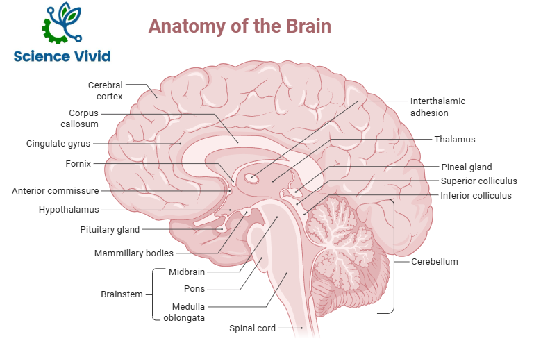
Fig: Anatomy of Brain
Meninges:
- Meninges are the three coverings around the brain & spine and help cushion, protect, and nourish the brain and spinal cord.
- Dura mater is a tough outermost layer made up of thick fibrous connective tissue
- The middle layer is called arachnoid mater, which attaches to the outer dura mater and has web-like linkages to the pia mater, which is the deepest layer.
- The fibrous trabeculae and blood vessels are found in the subarachnoid space. This area is where the CSF runs.
- The entire brain is covered with a very thin, translucent, but resilient covering called pia mater, which extends into all of the brain’s sulci and spinal cord.