Introduction:
The kidneys are two bean-shaped organs found in the back of the abdomen, one on each side of the spine. The nephron is the functional unit of the kidney, consisting of the glomerulus, proximal and distal tubules, and collecting duct. They are part of the urinary system and are in responsible of filtering waste products and toxins such as urea, creatinine, and uric acid, and excess fluid from the blood, regulating electrolyte balance.
It also synthesis hormones such as erythropoietin, 1,25 dihydroxy vitamin D, and renin hormones that regulate blood pressure and red blood cell production, and assisting in the maintenance of overall fluid and acid-base balance in the body.
Kidney disorders can lead to a variety of health concerns, such as chronic renal disease, kidney stones, infections, and cancer.
Renal function testing is essential in the therapy of patients with kidney disease. It is used to detect the existence of renal disease, assess the response of kidneys to treatment, and track the progress of renal illness.

Fig: Anatomy of kidney
kidney-related diseases:
- Chronic Kidney Disease (CKD): This is a long-term condition that affects the kidneys’ ability to filter blood and remove waste and excess fluids from the body.
- Acute Kidney Injury (AKI): This is a sudden and severe kidney injury that happens over a short period of time, often caused by dehydration, blood loss, or medication side effects.
- Kidney Stones: These are small, hard mineral deposits that form in the kidneys and can cause severe pain when passing through the urinary tract.
- Polycystic Kidney Disease (PKD): This is an inherited condition that causes numerous fluid-filled cysts to form in the kidneys, which can eventually lead to kidney failure.
- Glomerulonephritis: This is an inflammation of the kidneys’ filtering units (glomeruli) and can be caused by infections, immune disorders, or other conditions.
- Nephrotic Syndrome: This is a condition where the kidneys leak large amounts of protein into the urine, which can cause swelling, high blood pressure, and other complications.
- Renal Cell Carcinoma: This is a type of kidney cancer that develops in the cells of the renal tubules, which are the structures that filter blood in the kidneys.
- Pyelonephritis: This is a bacterial infection of the kidneys, which can cause fever, pain in the back or side, and other symptoms.
- Hemolytic Uremic Syndrome (HUS): This is a rare condition that can cause kidney failure and other serious health problems, often caused by E. coli or other bacteria.
- Alport Syndrome: This is an inherited condition that affects the kidneys’ filtering system and can cause kidney failure, hearing loss, and eye problems.
- Lupus Nephritis: This is a type of kidney inflammation that can occur in people with systemic lupus erythematosus (SLE), an autoimmune disease.
- IgA Nephropathy: This is a condition where antibodies build up in the kidneys’ filtering system and can cause inflammation and damage over time.
- Kidney Dysplasia: This is a condition where the kidneys do not develop properly during fetal development and can cause kidney failure later in life.
- Renal Artery Stenosis: This is a narrowing of the arteries that supply blood to the kidneys and can cause high blood pressure and kidney damage.
- Cystinosis: This is a rare genetic disorder that causes a buildup of cystine in the kidneys and other organs, which can lead to kidney failure and other health problems.
Renal function tests:
Renal function tests (RFT) are a collection of tests used to measure kidney (renal) function. The tests assess the amounts of numerous components in the blood, such as minerals, electrolytes, proteins, and glucose (sugar), creatinine, blood urea nitrogen (BUN), glomerular filtration rate (GFR), and electrolyte values such as sodium, potassium, and chloride to identify the current state of the kidneys. These tests can aid in the detection of kidney disease, the monitoring of kidney function in people with chronic renal disease or other illnesses, and the guidance of treatment decisions. Urine tests for protein, blood, and other compounds that can suggest kidney damage or disease may also be included in renal function testing.
When the kidneys aren’t working properly, waste products build up in the blood and fluid levels rise to dangerous levels, causing damage to the body or a potentially life-threatening situation. A variety of disorders and diseases can cause kidney injury. Diabetes and hypertension are the most frequent causes and risk factors for renal disease.
Specimen:
Renal function tests (RFTs) are blood and urine tests used to evaluate how well the kidneys are functioning. To obtain reliable and precise results, it is critical to follow any suggestions provided by the healthcare practitioner or lab regarding the time and preparation of RFT specimens.
Blood
A blood sample is taken from a vein in the arm to measure the levels of substances such as creatinine, blood urea nitrogen (BUN), and electrolytes, which can indicate kidney function. Fasting is not necessary for most RFTs, but some tests may require the patient to avoid food or drink for a certain period of time before the test.
- Blood urea nitrogen (BUN) test
- Creatinine test
- Glomerular filtration rate (GFR) test
- Serum electrolytes test (sodium, potassium, chloride, bicarbonate)
- Serum albumin test
- Serum calcium and phosphate test
Urine
A urine sample is collected to evaluate the presence of abnormal substances such as protein or blood, which may indicate kidney damage. The urine sample should be a clean-catch midstream sample, which involves collecting urine in a sterile container after cleaning the genital area. Some tests may require a 24-hour urine collection, in which all urine is collected over a 24-hour period and stored in a container for analysis.
- Urinalysis
- Urine protein test
- Urine creatinine test
- 24-hour urine collection
Components:
Blood Urea Nitrogen (BUN):
Urea, also known as BUN, is a nitrogen-containing component that is produced in the liver as a by-product of protein metabolism in the urea cycle. The kidneys eliminate around 85% of urea; the rest is eliminated through the gastrointestinal (GI) tract. Serum urea levels rise when renal clearance falls (as in acute and chronic renal failure/impairment). Urea levels can rise in circumstances other than kidney illness, such as upper GI haemorrhage, dehydration, catabolic states, and high protein diets. Urea levels may fall as a result of malnutrition, a low-protein diet, or severe liver illness. Serum creatinine is a better predictor of renal function than urea; nevertheless, urea rises early in renal failure.
When the BUN is elevated, the BUN: creatinine ratio can help distinguish between pre-renal and renal causes. The ratio is near to 20:1 in pre-renal disease, but closer to 10:1 in intrinsic renal disease. Upper GI haemorrhage has been linked to a very high BUN to creatinine ratio (often more than 30:1).
Creatinine:
Creatinine is a waste product that is produced by the muscles and filtered by the kidneys. It is used as a marker of kidney function in the renal function test. The renal function test is a blood test that measures the level of creatinine in the blood to assess how well the kidneys are functioning.
Glomerular filtration rate (GFR): (Clearance test)
A million small blood filtering units called nephrons are found within the kidneys. Blood is constantly filtered via a cluster of looping blood arteries called a glomerulus in each nephron, which enables water and tiny molecules to pass but traps blood cells, proteins like albumin, and bigger molecules.
Glomerular filtration rate (GFR), which is the rate in millilitres per minute at which chemicals in plasma are filtered through the glomerulus; in other words, the clearance of a substance from the blood, is the best test to assess glomerular function. A mature male’s GFR ranges between 90 and 120 mL per minute.
GFR can be determined using a simple blood test that measures creatinine levels. Creatinine is a by-product of protein digestion and the regular breakdown of muscle tissue. Aside from chronic renal disease, additional factors such as food, muscle mass, malnutrition, and other chronic conditions can also affect creatinine levels.
| Test | Normal Range | Interpretation |
| Blood Urea Nitrogen (BUN) | 7-20 mg/dL | Elevated levels indicate decreased kidney function |
| Creatinine | 0.6-1.3 mg/dL | Elevated levels indicate decreased kidney function |
| Estimated Glomerular Filtration Rate (eGFR) | >60 mL/min/1.73m2 | Lower eGFR indicates decreased kidney function |
| Serum Sodium | 135-145 mmol/L | Elevated levels indicate dehydration or kidney disease |
| Serum Potassium | 3.5-5.0 mmol/L | Elevated levels indicate kidney disease or dehydration |
| Serum Calcium | 8.5-10.2 mg/dL | Low levels indicate kidney disease or hypoparathyroidism |
| Serum Phosphorus | 2.5-4.5 mg/dL | Elevated levels indicate kidney disease or bone disease |
| Urine Protein | <150 mg/24 hours | Elevated levels indicate kidney disease or infection |
| Urine Albumin-to-Creatinine Ratio (ACR) | <30 mg/g | Elevated ACR indicates early kidney disease or risk of kidney disease |
| Kidney Biopsy | Diagnose kidney disease Examination of a small piece of kidney tissue under a microscope | Examination of a small piece of kidney tissue under a microscope |
| Renal Ultrasound | Evaluate kidney size, shape, and function | Ultrasound imaging of the kidneys |
Electrolytes: The kidneys are responsible for maintaining electrolyte balance in the body, so abnormal levels of electrolytes, such as potassium or sodium, can indicate kidney dysfunction.
Serum creatinine: This test measures the level of creatinine in the blood and is used to estimate GFR.
Albuminuria: This test measures the amount of albumin, a protein that can leak into the urine when the kidneys are damaged.
Cystatin C:
Cystatin C is a non-glycosylated low-molecular-weight serine protease inhibitor produced at a constant rate by all nucleated cells. It is found in body fluids and serum and freely filtered by the glomerulus but not secreted. It is reabsorbed by the tubules and completely metabolized there. Elevated levels of cystatin C in the blood can indicate reduced kidney function.
Renal ultrasonography: This imaging test uses sound waves to create pictures of the kidneys, which can help identify any structural abnormalities or blockages that may be causing kidney dysfunction.
Urinalysis:
Urinalysis involves detailed examination of kidney function through physical, chemical, and microscopic analysis of easily obtained urine samples. It is a cheap, productive test, and the readings can be obtained visually or through automated readings. Urinalysis should be employed as part of the initial investigation of patients.
| Test | Normal Range | Possible Causes of Abnormal Results |
| Colour | Pale yellow to amber | Dehydration, liver disease, bile duct obstruction, haematuria |
| Appearance | Clear | Infection, inflammation, kidney stones, dehydration |
| pH | 4.6-8.0 | Acidosis, alkalosis, respiratory or metabolic disorders |
| Specific Gravity | Dehydration, kidney disease, diabetes, liver disease | Dehydration, kidney disease, diabetes, liver disease |
| Glucose | Negative | Diabetes, pregnancy, liver disease |
| Protein | Negative | Kidney disease, urinary tract infection, dehydration |
| Ketones | Negative | Diabetes, low-carbohydrate diets, alcoholism |
| Bilirubin | Negative | Liver disease, bile duct obstruction, haemolytic anaemia |
| Urobilinogen | 0.2-1.0 mg/dL | Liver disease, haemolytic anaemia, hepatitis |
| Nitrites | Negative | Bacterial infection, urinary tract infection |
| Leukocyte Esterase | Negative | Bacterial infection, urinary tract infection |
| Microscopic Examination | ||
| Red Blood Cells (RBC) | 0-3 per high power field (HPF) | Kidney disease, bladder cancer, infection, kidney stones |
| White Blood Cells (WBC) | 0-5 per high power field (HPF) | Urinary tract infection, kidney disease, bladder cancer |
| Epithelial Cells | Few | Infection, inflammation, kidney stones Bacteria |
| Bacteria | None seen | Urinary tract infection, bacterial contamination |
| Crystals | None seen | Kidney stones, metabolic disorders, dehydration |
Immunological Marker (serological) test:
These immunological investigations can help in the diagnosis and management of various types of renal diseases, and may also help in monitoring the response to treatment. It is important to discuss the results of these tests with a healthcare professional who can provide further guidance and advice. Immunological investigations can be useful in the diagnosis and management of renal diseases. Some of the common immunological investigations performed in patients with renal disease include:
Antinuclear Antibody (ANA) Test:
This test detects the presence of autoantibodies directed against the nuclei of cells. ANA is used to diagnose and monitor systemic lupus erythematosus (SLE), which is a common cause of renal disease.
Anti-double-stranded DNA (Anti-dsDNA) Test:
This test measures the level of anti-dsDNA antibodies in the blood. Elevated levels of anti-dsDNA antibodies are found in patients with SLE and can indicate an increased risk of renal disease.
Anti-glomerular Basement Membrane (Anti-GBM) Antibody Test:
This test detects the presence of anti-GBM antibodies in the blood. Elevated levels of anti-GBM antibodies are found in patients with Goodpasture’s syndrome, a rare autoimmune disorder that can cause rapidly progressive glomerulonephritis (inflammation of the glomeruli in the kidneys).
Complement Component 3 (C3) and Complement Component 4 (C4) Test:
This test measures the levels of complement proteins C3 and C4 in the blood. Decreased levels of C3 and C4 are found in patients with SLE and other autoimmune disorders.
Antineutrophil Cytoplasmic Antibodies (ANCA) Test:
This test detects the presence of ANCA antibodies in the blood. Elevated levels of ANCA antibodies are found in patients with autoimmune vasculitis, including granulomatosis with polyangiitis (GPA) and microscopic polyangiitis (MPA), which can cause renal disease.
Rheumatoid Factor (RF) Test:
This test measures the level of RF antibodies in the blood. Elevated levels of RF antibodies are found in patients with rheumatoid arthritis, which can cause glomerulonephritis and renal disease.
Molecular diagnosis:
Molecular testing is not commonly used for routine evaluation of renal function. However, molecular tests can be useful in certain cases where a genetic disorder is suspected as the underlying cause of kidney disease.
For example, genetic testing can be used to diagnose autosomal dominant polycystic kidney disease (ADPKD), a genetic disorder that causes the growth of numerous cysts in the kidneys, leading to kidney failure. ADPKD is caused by mutations in the PKD1 or PKD2 genes, and molecular testing can identify these mutations in affected individuals and their family members.
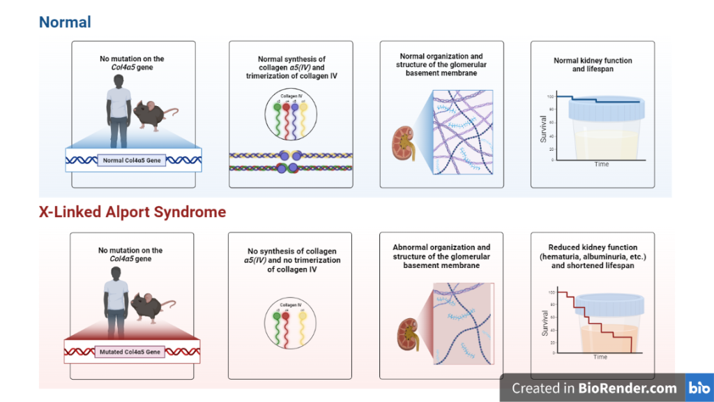
Another example is Alport syndrome, a genetic disorder that affects the kidneys and can lead to kidney failure. Alport syndrome is caused by mutations in genes encoding for type IV collagen, a component of the glomerular basement membrane in the kidneys. Molecular testing can identify these mutations in affected individuals and their family members.
In addition, molecular testing can be used to assess the risk of developing certain types of kidney cancer, such as von Hippel-Lindau (VHL) syndrome, which is caused by mutations in the VHL gene.
Overall, molecular testing can be a useful tool in the diagnosis and management of certain genetic kidney disorders, but it is not typically used for routine assessment of renal function.

Fig: X-Linked Alport Syndrome Pathology
| Test | Indications | Examples |
| Sanger sequencing | Disorders with minimal locus heterogeneity | Fabry disease (GLA), Denys–Drash (WT1), cystinosis (CTNS) |
| CGH/SNP array, MLPA | Large CNVs suspected | CAKUT, aHUS (CFH, CFHR), nephronophthisis (NPHP1) |
| Targeted phenotype- associated gene panel Targeted ES (virtual gene panel) | Disorders with locus heterogeneity Disorders with overlapping phenotypes Disorders associated with genes from common pathway | SRNS Hereditary tubulopathies, Complement-related disorders |
| Exome sequencing (ES) | Phenotype indistinct and underlying cause unknown Second-tier test after gene panel testing | Unexplained kidney failure |
| Genome sequencing (GS) | Due to high costs, interpretation challenges and long analytical period, currently only used in research for cases still unsolved after ES Emerging clinical use | ADPKD (PKD1) |
Table: Various molecular testing modalities and their existing nephrology indications
Different testing modalities and their current indications in nephrology:
| Testing Modality | Indications |
| Urine analysis | Detection of blood, protein, and other substances in the urine to diagnose glomerulonephritis, urinary tract infections, and nephrotic syndrome |
| Blood tests (serum creatinine, BUN, eGFR) | Assessment of kidney function and detection of abnormalities in the blood that may indicate kidney disease |
| Kidney biopsy | Examination of kidney tissue under a microscope to diagnose various kidney diseases such as glomerulonephritis, vasculitis, and nephrotic syndrom |
| Imaging tests (ultrasound, CT scan, MRI) | Diagnosis of kidney stones, tumours, and other abnormalities in the kidneys |
| Dialysis adequacy testing | Assessment of the effectiveness of dialysis treatment in removing waste and excess fluid from the body |
| Genetic testing | Identification of inherited kidney diseases such as polycystic kidney disease (PKD) and Alport syndrome |
References:
- Henry O. Ogedegbe, PhD, BB(ASCP)SC, CLS(NCA), Renal Function Tests: A Clinical Laboratory Perspective, Laboratory Medicine, Volume 38, Issue 5, May 2007, Pages 295–304.
- Ogedegbe, H.O., 2007. Renal function tests: A clinical laboratory perspective. Laboratory Medicine, 38(5), pp.295-304.
- Bishop ML, Fody EP, Schoeff L. Clinical Chemistry, Principles, Procedures, Correlations. 5th Edition. Shrzynski CJ, Wu AHB. Lippincott Williams and Wilkins; 2005:517–537.
- Gounden, V., Bhatt, H. and Jialal, I., 2018. Renal function tests.
- Winearls, Christopher G., Jo H. M. Berden, and Jack F. M. Wetzels (ed.), ‘Immunological investigation of the patient with renal disease’, in Neil N. Turner and others (eds), Oxford Textbook of Clinical Nephrology: Three-Volume Pack, 4 edn (Oxford, 2015; online edn, Oxford Academic, 1 June 2019).
- Ronco, C. Acute kidney injury: from clinical to molecular diagnosis. Crit Care 20, 201 (2016).

