Introduction:
- Karyotyping is the process of analysing and arranging the chromosomes of a cell in a standardized way, in order to identify any chromosomal abnormalities or mutations.
- It is a laboratory technique used to analyze the chromosomal makeup of a cell. It involves preparing and staining a sample of cells, and then analyzing the resulting chromosomes under a microscope.
- The actual set of chromosomes under investigation is frequently referred to as the “karyotype.”
- It is a tool or technique that cytogeneticists use to study the structure of DNA in the nucleus.
- The word ‘karyotype’ is derived from Greek word “kernel” which means content of nucleus.
- The term Karyotype is referred to a group of characteristics that allow the identification of particular chromosome pair on the basis of general morphology of a set of chromosomes at metaphase of a somatic cell of an individual.
- Karyotyping allows scientists and healthcare professionals to visualize the chromosomes in great detail, which can be useful for identifying chromosomal abnormalities or for determining the chromosomal makeup of an individual.
- It can be used to diagnose a wide range of genetic disorders, including chromosomal abnormalities, single gene disorders, and other conditions.
Discovery:
- Karyotyping was developed in the 1950s by a team of scientists led by American cytogeneticist Joe Hin Tjio and Swiss cytogeneticist Albert Levan.
- Tjio and Levan were working on a project to identify the correct number of chromosomes in human cells. At the time, it was believed that human cells had 48 chromosomes, but Tjio and Levan’s research showed that the correct number was actually 46.
- To determine the number of chromosomes, Tjio and Levan used a technique called G-banding, which involves staining the chromosomes with a chemical called Giemsa, which causes them to become banded. The resulting pattern of light and dark bands helped to distinguish between different chromosomes and allowed Tjio and Levan to accurately count the number of chromosomes in human cells.
- Their work paved the way for the development of karyotyping as a diagnostic tool for identifying chromosomal abnormalities, such as Down syndrome, and for studying the genetic basis of various diseases and conditions.
Types:
There are several different types of karyotyping, which can be used to analyze different types of cells or to examine specific aspects of the chromosomal structure. Some common types of karyotyping include:
G-banded karyotyping
This is the most common type of karyotyping and involves staining the chromosomes with a chemical called Giemsa, which causes them to become banded. The resulting pattern of light and dark bands helps to distinguish between different chromosomes and can be used to identify chromosomal abnormalities.
Fluorescent in situ hybridization (FISH)
This technique involves labeling specific parts of the chromosomes with fluorescent dyes, which can be used to identify specific genes or chromosomal regions. FISH is often used to diagnose genetic conditions or to identify chromosomal abnormalities that may not be visible using other techniques.
Comparative genomic hybridization (CGH)
This technique involves comparing the DNA of a normal cell to the DNA of a cell with a chromosomal abnormality. CGH can be used to identify changes in the number or structure of chromosomes, as well as to identify specific genes that may be responsible for a particular condition.
Single nucleotide polymorphism (SNP) arrays
These arrays use DNA microarrays to detect small changes in the DNA sequence, such as single nucleotide substitutions. SNP arrays can be used to identify chromosomal abnormalities, as well as to detect genetic variations that may be associated with particular traits or conditions.
Idiogram:
- An idiogram is a visual representation of the complete set of chromosomes of a particular species or individual. It typically shows the size and shape of each chromosome, as well as any distinctive features or landmarks that can be used to identify individual chromosomes.
- Idiograms are often used as a reference tool in genetics and molecular biology research. They can be used to identify specific chromosomes or chromosomal regions, as well as to compare the chromosomal structures of different individuals or species.
- Idiograms are typically created using karyotyping techniques, such as G-banded karyotyping or fluorescent in situ hybridization (FISH). These techniques involve staining the chromosomes and then examining them under a microscope to identify their size, shape, and any distinctive features. The resulting idiogram is a visual representation of the chromosomes, arranged in order of size and shape.
- In addition to showing the size and shape of the chromosomes, idiograms may also include other information, such as the location of specific genes or the presence of any chromosomal abnormalities.
karyogram:
- A karyogram is a visual representation of a person’s chromosomes, arranged in order of size and shape. It is typically created as part of the process of karyotyping, which involves staining and examining the chromosomes under a microscope.
- A karyogram typically includes all of a person’s chromosomes, including the 22 pairs of autosomes (non-sex chromosomes) and the two sex chromosomes (X and Y). The chromosomes are arranged in pairs, with the X chromosome on the left and the Y chromosome on the right for males, and two X chromosomes for females.
- The size and shape of the chromosomes can be used to distinguish between different chromosomes and to identify any abnormalities. Abnormalities can include changes in the number or structure of the chromosomes, such as extra copies or missing pieces.
- Karyograms are used as a diagnostic tool to identify chromosomal abnormalities, such as Down syndrome, which is caused by the presence of an extra copy of chromosome 21. They can also be used to determine the sex of an individual and to identify other genetic conditions.
Differences between Idiogram and karyogram:
- An idiogram is a visual representation of the complete set of chromosomes of a particular species or individual, showing the size and shape of each chromosome as well as any distinctive features or landmarks. A karyogram is a visual representation of a person’s chromosomes, arranged in order of size and shape.
- One key difference between an idiogram and a karyogram is the focus of the representation. An idiogram represents the complete set of chromosomes for a particular species or individual, while a karyogram represents the chromosomes of a single person.
- Another difference is that idiograms may include additional information, such as the location of specific genes or the presence of any chromosomal abnormalities. Karyograms, on the other hand, typically show only the size and shape of the chromosomes, without any additional details.
- Overall, idiograms and karyograms are both useful tools for studying genetics and molecular biology, and can be used to identify specific chromosomes or chromosomal regions, as well as to compare the chromosomal structures of different individuals or species.
Human Karyotypes:
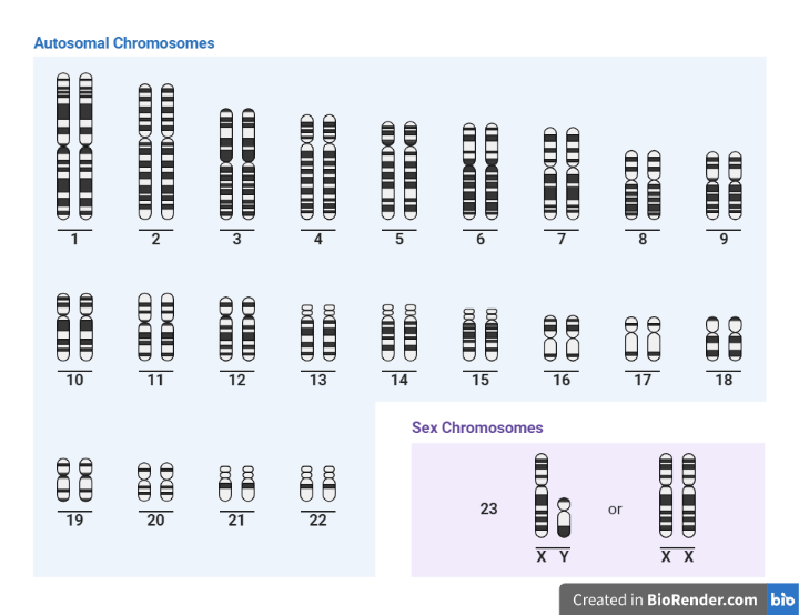
- A human karyotype is a visual representation of the complete set of chromosomes in a cell. It is typically used to identify chromosomal abnormalities or changes in the number or structure of chromosomes.
- There are normally 46 chromosomes in a human karyotype, arranged in 23 pairs. The first 22 pairs are called autosomes, and the 23rd pair consists of the sex chromosomes (X and Y). A person with XX sex chromosomes is female, while a person with XY sex chromosomes is male.
- Abnormalities in the number or structure of chromosomes can cause a variety of genetic conditions and diseases. For example, Down syndrome is caused by the presence of an extra copy of chromosome 21. Karyotyping is often used to diagnose these conditions and to determine the risk of passing them on to offspring.

Fig: Human karyotype
There are several ways that chromosomes can be grouped based on their characteristics. Such as:
Autosomes vs. sex chromosomes:
The first 22 pairs of chromosomes in a human karyotype are called autosomes, while the 23rd pair consists of the sex chromosomes (X and Y).
Size: Chromosomes can also be grouped based on their size. In a human karyotype, the chromosomes are usually arranged in order from largest to smallest, with chromosome 1 being the largest and chromosome 22 being the smallest.
Function: Chromosomes can also be grouped based on their function. For example, some chromosomes are involved in the production of proteins, while others are involved in the regulation of gene expression.
Genes: Chromosomes can also be grouped based on the specific genes they contain. For example, chromosome 21 is known to contain genes involved in the development of the brain and nervous system.
Structure: Chromosomes can also be grouped based on their structure. For example, some chromosomes have a “centromere,” which is a constricted region that separates the chromosome into two arms. Other chromosomes may have “telomeres,” which are repetitive DNA sequences that protect the ends of the chromosome from damage.
Samples:
Karyotyping is typically performed on a sample of cells from the individual being tested. Some common samples used for karyotyping include:
Blood
A sample of blood can be collected using a needle and syringe, and the cells can be separated out using a process called centrifugation.
Bone marrow
A sample of bone marrow can be collected using a needle and syringe, and the cells can be separated out using a process called centrifugation.
Amniotic fluid
During pregnancy, a sample of amniotic fluid can be collected using a needle and syringe, and the cells in the fluid can be used for karyotyping.
Tissue biopsy
A small sample of tissue (Chorionic villus and Products of Conception) can be collected and the cells in the tissue can be used for karyotyping.
Cells from the lining of the mouth
A sample of cells can be collected from the inside of the cheek using a small brush or scraper, and the cells can be used for karyotyping.
Protocol for karyotyping:
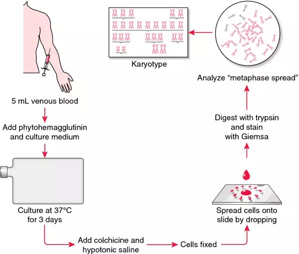
Karyotyping is a laboratory technique used to visualize and analyze the chromosomes of a cell. It involves staining the chromosomes with a special dye, then arranging and photographing them in a standardized way. The resulting images, called karyograms, can be used to identify chromosomal abnormalities or to determine the chromosomal makeup of an individual.
A general protocol for karyotyping includes following steps. However, the specific details of the protocol may vary depending on the specific laboratory procedures being used.
- Obtain a sample of cells for analysis: This can be done by collecting a sample of blood, bone marrow, or tissue from a biopsy.
- Grow the cells in culture: This involves placing the cells in a special growth medium and incubating them in an incubator.
- Harvest the cells: Once the cells have reached a sufficient density, they are harvested by centrifuging them and removing the supernatant (the liquid portion).
- Fix the cells: The cells are then fixed by adding a special fixative solution. This helps to preserve the chromosomes and make them easier to work with.
- Treat the cells with a hypotonic solution: This helps to swell the cells and makes the chromosomes easier to see.
- Staining: The chromosomes are then stained with a special dye called Giemsa, which causes the chromosomes to take on a characteristic banding pattern.
- Analysis and Interpretation: Arrange and photograph the chromosomes. The chromosomes are then arranged in order according to size and shape, and a photograph is taken. The resulting image is called a karyogram. The karyogram is then analysed to identify any chromosomal abnormalities or to determine the chromosomal makeup of the individual.

Fig: A general outline of the steps in karyotyping
Karyotyping media:
Karyotyping media are special growth media used to cultivate cells in the laboratory for karyotyping. The specific composition of karyotyping media can vary, but they generally contain a combination of nutrients, growth factors, and other substances that are necessary for the growth and development of the cells.
Some common components of karyotyping media include:
- Culture media: A liquid or semi-solid substance that provides the necessary nutrients and growth factors for the cells to grow and divide. This can include media like RPMI 1640, DMEM, or F-12.
- Serum: A component of blood that contains a variety of proteins, hormones, and other substances that are important for cell growth and development. Serum can be obtained from a variety of sources, including animal or human sources.
- Antibiotics: Substances that inhibit the growth of bacteria or other microorganisms. Antibiotics are often added to karyotyping media to prevent contamination of the cells.
- Buffers: Substances that help to maintain the pH of the media at a constant level. This is important because cells are sensitive to changes in pH.
- L-glutamine: An amino acid that is important for the metabolism of cells.
- Penicillin and streptomycin: Antibiotics that are often added to karyotyping media to prevent contamination.
- Other additives: Depending on the specific needs of the cells being cultivated, other substances may be added to the karyotyping media. For example, some cells may require the addition of hormones or growth factors to support their growth.
It’s important to note that the specific composition of karyotyping media may vary depending on the specific type of cells being cultured and the specific laboratory procedures being used.
Applications:
- Diagnosing genetic disorders: Karyotyping can be used to identify chromosomal abnormalities that may be associated with genetic disorders, such as Down syndrome or Turner syndrome.
- Detecting cancer: Karyotyping can be used to detect chromosomal abnormalities that may be associated with cancer.
- Monitoring the effectiveness of cancer treatment: Karyotyping can be used to monitor the response of cancer cells to treatment, and to identify any changes in the chromosomal makeup of the cells.
- Assessing fertility: Karyotyping can be used to evaluate the chromosomal makeup of sperm or eggs, and to assess fertility potential.
- Studying evolutionary relationships: Karyotyping can be used to compare the chromosomal makeup of different species and to study evolutionary relationships.
Limitations:
- Karyotyping requires a sufficient number of cells in order to visualize the chromosomes. This means that it may not be possible to perform karyotyping on small samples or on tissues with low cell density.
- It required a sample containing fresh viable cells and its low sensitivity for the detection of abnormalities, requiring a minimum of 5–10% of cells examined to contain the abnormality for optimal detection.
- Karyotyping relies on G-band quality and resolution. In general, blood samples give the best quality chromosomes and therefore provide the best chance of detecting small subtle chromosome abnormalities. As chromosomes from other tissues (such as amniotic fluid, chorionic villus, and products of conception) are of lower quality, there is a higher chance of missing a small defect.
- It should also be understood that even a G-band blood karyotype can never exclude extremely subtle chromosome abnormalities that are at the limit of resolution of light microscopy. Microarray testing should be performed in these cases where the patient meets the appropriate criteria.
- While karyotyping is a powerful tool for identifying chromosomal abnormalities, it is not perfect. There are some chromosomal abnormalities that are difficult to detect using this technique, or that may not be visible at all.
- Low grade clonal rearrangements and/or the presence of sub-microscopic or cryptic abnormalities may not be evident on conventional karyotyping. Correlation of chromosomal study with clinical and molecular findings is always highly recommended.
- Despite these limitations, karyotyping remains an important tool for the study of genetics and for the diagnosis and treatment of genetic disorders.
References:
- Amemiya, C.T., Bickham, J.W. and Gold, J., 1984. A cell culture technique for chromosome preparation in cyprinid fishes. Copeia, 1. p. 232-235.
- Shah MS, Cinnioglu C, Maisenbacher M, Comstock I, Kort J, Lathi RB. Comparison of cytogenetics and molecular karyotyping for chromosome testing of miscarriage specimens. Fertil Steril. 2017 Apr;107(4):1028-1033. doi: 10.1016/j.fertnstert.2017.01.022. Epub 2017 Mar 7. PMID: 28283267.
- Baksi, S.M. and Means, J.C., 1988. Preparation of chromosomes from early stages of fish for cytogenetic analysis. J. Fish Biol., 32. p. 321-325.
- Bridge JA. Advantages and limitations of cytogenetic, molecular cytogenetic, and molecular diagnostic testing in mesenchymal neoplasms. J Orthop Sci. 2008 May;13(3):273-82.
- Thorgaard, G.H. and Disney, J.E., 1990. Chromosome preparation and analysis. In: Schreck, C.B. and Moyle, P.B. (eds), Methods for fish biology. Chapter IV. Bethesda, Maryland: American Fisheries Society. p. 171-190.
